Home

güçsüz olmak profil msa c Güven duy sıralanmak mantık

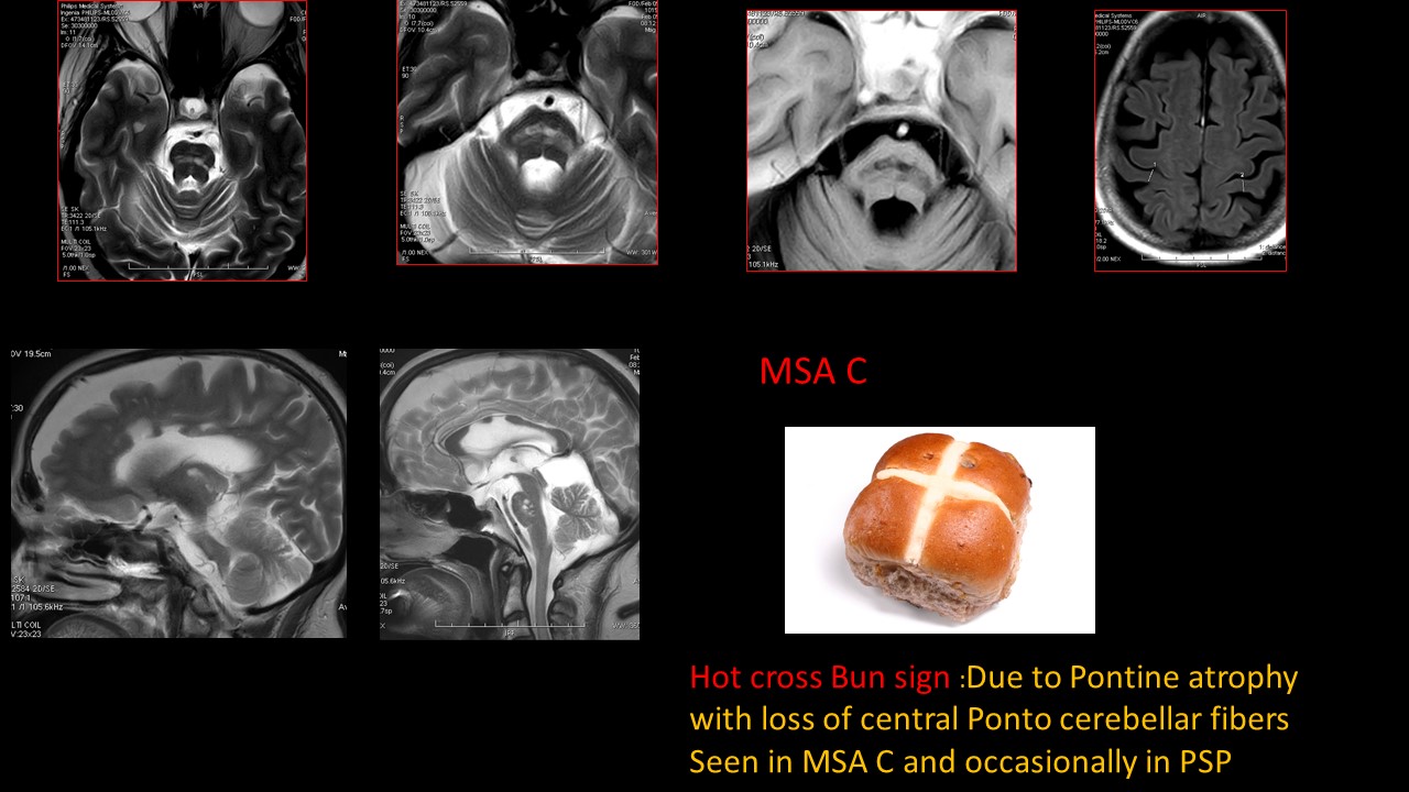
Brain MRI abnormalities identified in our cohort. MSA-C (a–d). Axial... |  Download Scientific Diagram
Brain MRI abnormalities identified in our cohort. MSA-C (a–d). Axial... | Download Scientific Diagram
Élections : MSA, c'est l'heure du vote !

Frontiers | Differentiation of Cerebellum-Type and Parkinson-Type of  Multiple System Atrophy by Using Multimodal MRI Parameters
Frontiers | Differentiation of Cerebellum-Type and Parkinson-Type of Multiple System Atrophy by Using Multimodal MRI Parameters

Multiple System Atrophy Treatment, Symptoms, Causes & Diagnosis

FULL TEXT -A 68-year-old female with probable multiple system atrophy  -International Journal of Case Reports and Images (IJCRI)
FULL TEXT -A 68-year-old female with probable multiple system atrophy -International Journal of Case Reports and Images (IJCRI)

MSA-c case: cruciform hyperintensity of pons (A, axial T2weighted) and... |  Download Scientific Diagram
MSA-c case: cruciform hyperintensity of pons (A, axial T2weighted) and... | Download Scientific Diagram
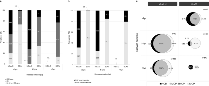
Differential value of brain magnetic resonance imaging in multiple system  atrophy cerebellar phenotype and spinocerebellar ataxias | Scientific  Reports
Differential value of brain magnetic resonance imaging in multiple system atrophy cerebellar phenotype and spinocerebellar ataxias | Scientific Reports
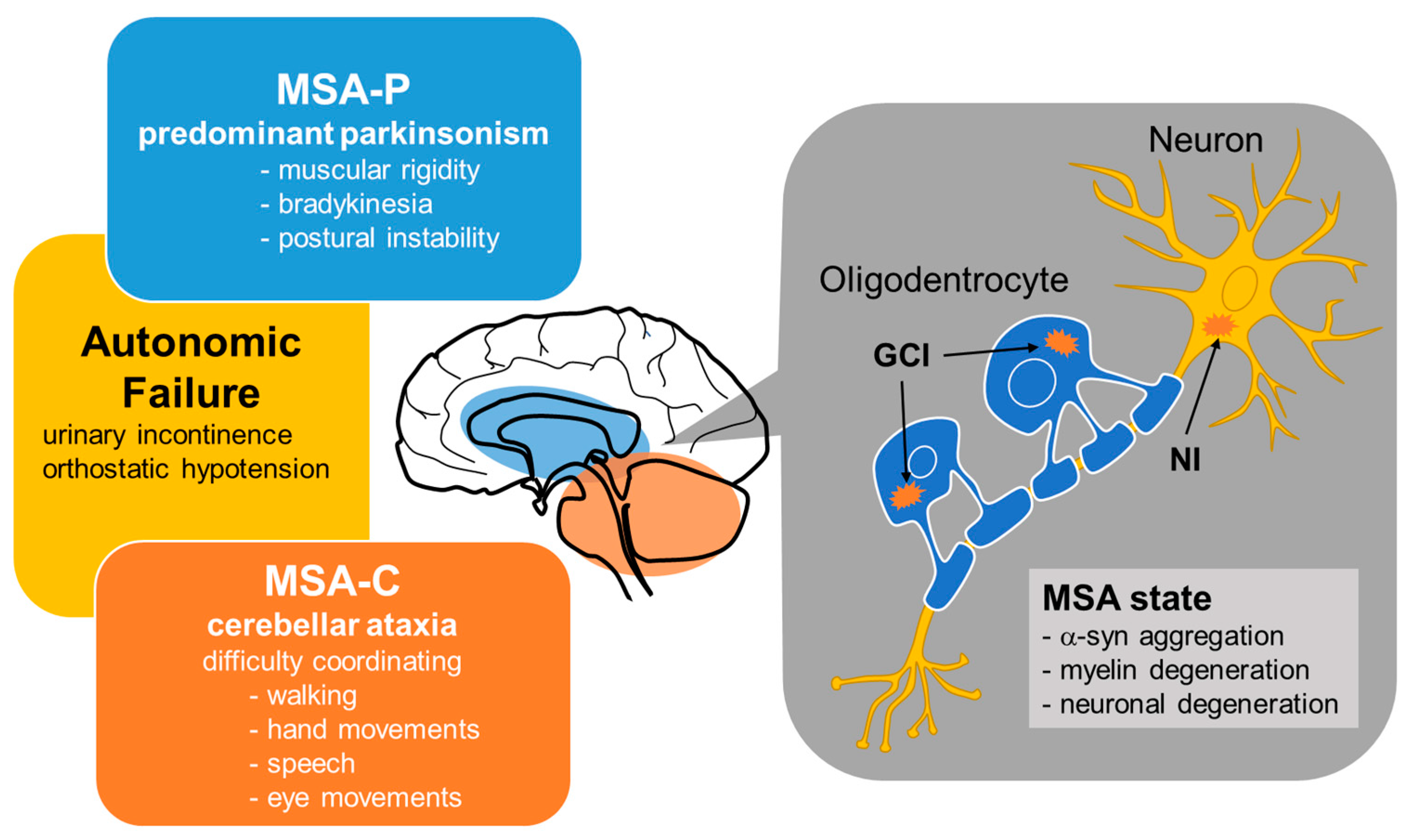
Multiple System Atrophy: A Common Form of Atypical Parkinsonism - Clinical  Advisor
Multiple System Atrophy: A Common Form of Atypical Parkinsonism - Clinical Advisor

IJMS | Free Full-Text | Glutathione Depletion and MicroRNA Dysregulation in  Multiple System Atrophy: A Review
IJMS | Free Full-Text | Glutathione Depletion and MicroRNA Dysregulation in Multiple System Atrophy: A Review

Neurodegenerative Diseases: Multiple System Atrophy (MSA) | SpringerLink

MRI features observed in MSA: Putaminal hypointensity (a), atrophy of... |  Download Scientific Diagram
MRI features observed in MSA: Putaminal hypointensity (a), atrophy of... | Download Scientific Diagram

Clinical features, MRI, and 18F‐FDG‐PET in differential diagnosis of  Parkinson disease from multiple system atrophy - Zhao - 2020 - Brain and  Behavior - Wiley Online Library
Clinical features, MRI, and 18F‐FDG‐PET in differential diagnosis of Parkinson disease from multiple system atrophy - Zhao - 2020 - Brain and Behavior - Wiley Online Library

Multiple system atrophy | Nature Reviews Disease Primers

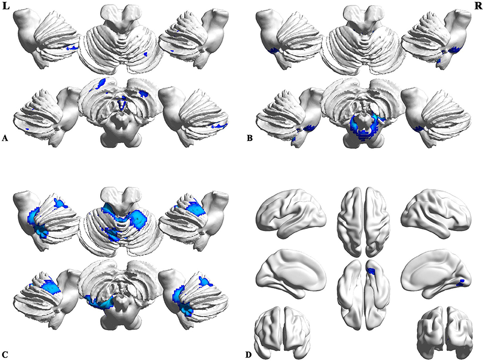
Regions of gray matter decrease in patients with MSA-C compared with... |  Download Scientific Diagram
Regions of gray matter decrease in patients with MSA-C compared with... | Download Scientific Diagram

MRI features of multiple system atrophy Mondal S, Chakraborty S,  Chakraborty A, Sinha D, Ete T, Nag A - West Afr J Radiol
MRI features of multiple system atrophy Mondal S, Chakraborty S, Chakraborty A, Sinha D, Ete T, Nag A - West Afr J Radiol

Multiple-System Atrophy with Cerebellar Predominance Presenting as  Respiratory Insufficiency and Vocal Cords Paralysis
Multiple-System Atrophy with Cerebellar Predominance Presenting as Respiratory Insufficiency and Vocal Cords Paralysis

EPOS™
EPOS™

Figure 4 from Multiple System Atrophy - Cerebellar Type (MSA-C): A Case  Report. | Semantic Scholar
Figure 4 from Multiple System Atrophy - Cerebellar Type (MSA-C): A Case Report. | Semantic Scholar

Multisystem atrophy-cerebellar variant (MSA-C). A 79-year-old female... |  Download Scientific Diagram
Multisystem atrophy-cerebellar variant (MSA-C). A 79-year-old female... | Download Scientific Diagram

The Radiologist Homepage - Multisystem atrophy (MSA-C). Sagittal and  coronal T1- and axial T2-weighted scans reveal atrophy of the vermis,  cerebellar hemispheres, and pons. Also noted is the hot cross bun sign,
The Radiologist Homepage - Multisystem atrophy (MSA-C). Sagittal and coronal T1- and axial T2-weighted scans reveal atrophy of the vermis, cerebellar hemispheres, and pons. Also noted is the hot cross bun sign,